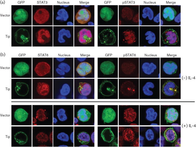
Fig. 2.
Tip induces nuclear translocation of STAT6. Jurkat T-cells were electroporated with plasmids encoding GFP (vector) or GFP–Tip together with pVR/STAT3 or STAT6. Twenty-four hours after electroporation, localization of STAT3 (a) and STAT6 (b) was examined by confocal microscopy after staining with anti-STAT or anti-phosphoSTAT antibodes (red). TO-PRO-3 staining was used to visualize the nucleus (blue). Nuclear translocation of STAT6 was also examined in the absence (−) or presence (+) of IL-4 treatment (100 ng ml−1, 15 min).

