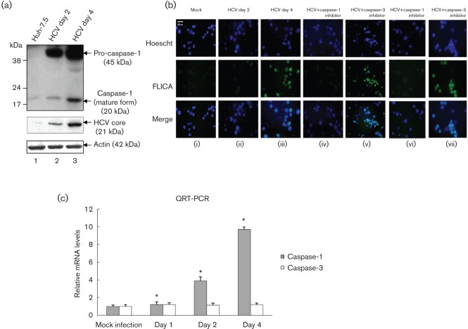
Fig. 2.
Activation of caspase-1 by HCV. (a) Mock-infected and HCV-infected Huh7.5 cells were serum starved for 4 h and cellular lysates were subjected to SDS-PAGE followed by Western blot analysis using caspase-1 antibody. Lane 1, Huh7.5 lysates; lanes 2 and 3, lysates from Huh7.5 cells infected with HCV. Bottom panels represent the expression of HCV core and actin controls. (b) FLICA assay. Mock-infected and HCV-infected cells were serum starved for 4 h and treated with 50 µM caspase-1 inhibitor (z-VAD-fmk) for 1 h (iv), and 2 h (vi) or 100 µM caspase-3 inhibitor (DEVD) for 1 h (v), and 2 h (vii). The cells were incubated with the FLICA reagent and Hoescht nuclear stain for 1 h, before being visualized via fluorescence microscopy. (c) Total cellular RNA from mock-infected and HCV-infected cells were subjected to QRT-PCR using caspase-1-specific primers. The data shown here represent the means+sd of at least three independent experiments performed in triplicate. *, P<0.05 compared with mock-infected control cells.

