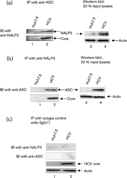
Fig. 6.
Interaction of ASC with NALP3. Mock-infected and HCV-infected cells were cultured for 4 days and serum starved for 4 h. Equal amounts (500 µg) of cellular lysates from mock-infected and HCV-infected cells were immunoprecipitated with anti-ASC (a), anti-NALP3 (b) or isotype control antibodies (c), and immunoblotted with anti-NALP3 or anti-ASC antibodies. Lane 1, mock-infected lysates; lane 2, HCV-infected lysates. Bottom panels represent the expression of HCV core protein and actin protein loading controls. Lane 4 in (a) and (b) represent 20 % input lysates from mock-infected and HCV-infected cells.

