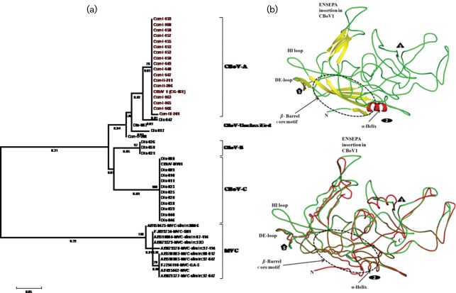
Fig. 3.
(a) Genetic diversity of structural proteins among CBoV variants found in diseased (Dis-disease) and healthy (Con-control) dogs. For comparison, genetic diversity among MVC variants was included in the analysis (lower branch of the tree); and (b) comparative secondary structure of the capsid protein of CBoV variants was used to decipher the structural changes caused by insertion of six amino acid residues. In the ribbon diagram of CBoV1, the secondary structure elements (β-strand in yellow, helix in red and loop in green) are coloured differently. The icosahedral symmetry axes are represented as oval, triangle and pentagon. In the coil representation of CBoV capsid structure the CBoV-A and -C are shown as red and green, respectively. Bars, represent 0.05 substitutions per amino acid site.

