Figure 3.

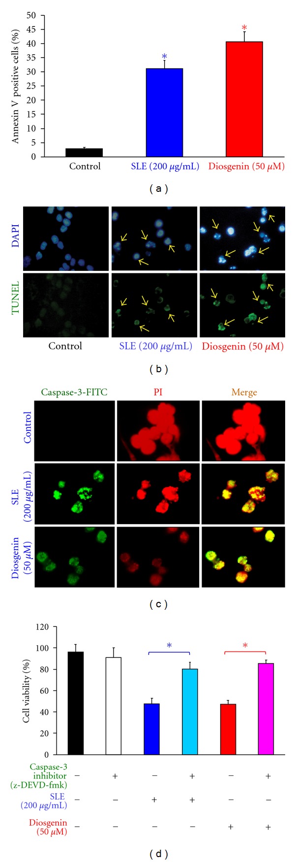
SLE- and diosgenin-induced apoptosis and caspase-3 activation in WEHI-3 cells. Cells were treated with 200 μg/mL of SLE or 50 μM of diosgenin for 12 h. (a) Annexin V/PI analysis was determined by flow cytometric assay. Apoptotic cell population (Annexin V positive cells) was quantified as described in materials and methods. Data are presented as the mean ± S.E.M. of three independent experiments.*, P < 0.05, significantly different compared with control treatment. Cells were treated with 200 μg/mL of SLE or 50 μM of diosgenin for 48 h. (b) DAPI/TUNEL analysis and (c) caspase-3 protein location were determined by immunostaining and photographed by fluorescence microscopic systems as described in materials and methods (400X) (↑DNA fragmentation). (d) Cells were pretreated with specific inhibitor of caspases-3 (z-DEVD-fmk) for 1 h after exposure to SLE (200 μg/mL) or diosgenin (50 μM) for 48 h exposure. The cells were collected to determine the percentage of viable cells. Data are presented as the mean ± S.E.M. of three independent experiments. *, P < 0.05, significantly different compared with SLE-treated cells.
