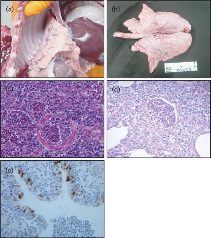
Fig. 1.
(a, b) Cranioventral consolidation. Note the distinctive chessboard-like distribution over the lung in (b). (c, d) Histopathology of the case shown in (a). Medium-sized bronchioles were lined with immature flat epithelial cells (c), whilst small bronchioles showed necrosis and sloughed-off epithelium (d). The lumen was filled with detached necrotic epithelial cells and neutrophils. (e) Immunohistochemical staining showed IAV antigen on the epithelial cells in bronchioles that showed slight inflammatory changes and a few scattered mononuclear cells within the alveoli. Magnification, ×40 (c–e).

