Figure 1:

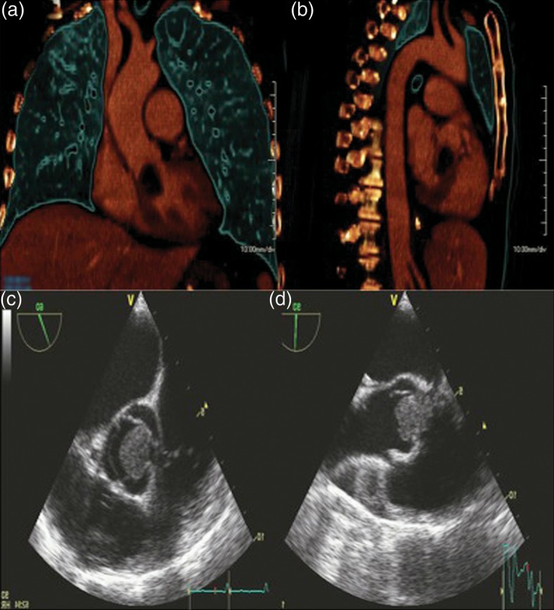
3D reconstruction of coronal and sagittal views of the chest demonstrating the large cardiac Aspergilloma. Parasternal long- and short-axis views on transthoracic echocardiography showing LVOT mass.

3D reconstruction of coronal and sagittal views of the chest demonstrating the large cardiac Aspergilloma. Parasternal long- and short-axis views on transthoracic echocardiography showing LVOT mass.