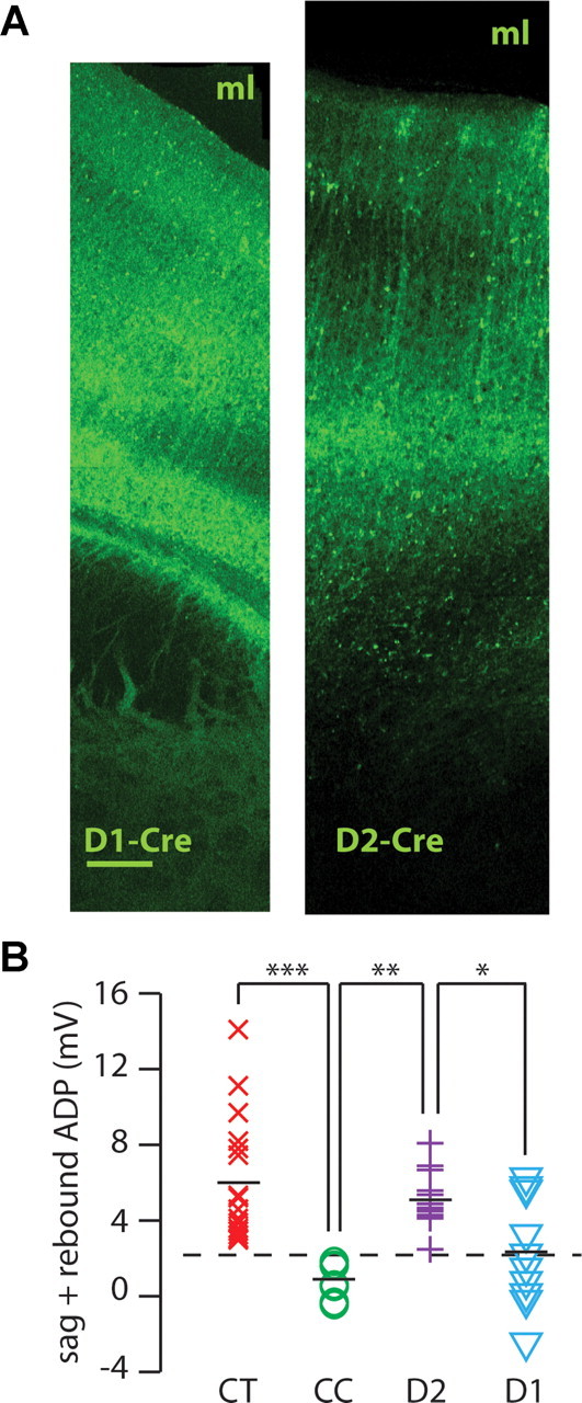
Figure 3.

D2Rs are selectively expressed in type A pyramidal neurons which can be distinguished using the h-current. A, Low-power confocal images of infralimbic cortex showing the pattern of fluorescence in Drd1-Cre (D1-Cre) and Drd2-Cre (D2-Cre) transgenic mice injected with virus to drive Cre-dependent expression of ChR2-YFP. The corpus callosum and midline lie below and above both images, respectively. ml, midline. Scale bar, 0.1 mm (both images are to the same scale). B, The amount of h-current (measured as above) in identified corticothalamic (CT, n = 18), corticocortical (CC, n = 8), D2R-expressing (D2, n = 14), or D1R-expressing (D1, n = 10) pyramidal neurons in layer V of mPFC. The dotted line indicates the threshold that separates the distributions of h-current from CT and CC neurons. *p < 0.05, **p < 0.01.
