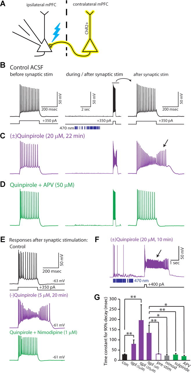
Figure 4.

Synaptic stimulation unmasks a novel D2R-mediated afterdepolarization in specific layer V pyramidal neurons. A, Experimental design. We recorded from ChR2-negative layer V neurons while stimulating ChR2-positive axons from the contralateral mPFC with trains of light flashes (470 nm, 2.5 ms, ∼2 mW). B, Responses of a type A layer V pyramidal neuron to current injection before (left) and immediately following (middle and right) optogenetic stimulation of synaptic inputs. Blue bars indicate the times of light flashes. C, Before synaptic stimulation, no quinpirole-induced afterdepolarization is observed; however, the same current injection elicits a marked afterdepolarization (along with spike height rundown) following weak synaptic stimulation. D, The quinpirole-induced afterdepolarization is eliminated by the addition of AP5. E, Lower doses of quinpirole (5 μm) also induce an afterdepolarization following synaptic stimulation, which can be blocked by nimodipine (1 μm). F, Recording from a type A neuron showing a prolonged quinpirole-induced afterdepolarization following synaptic stimulation. G, Average time constants for the membrane potential to return to baseline following a depolarizing current pulse (300–400 pA, 250–500 ms) delivered immediately following the pattern of synaptic stimulation shown above. Data are shown for control conditions (black; n = 12), quinpirole (purple; 5 μm, n = 7; 20 μm, n = 6), quinpirole in the absence of synaptic stimulation (hollow purple bar; 5 μm, n = 6), nimodipine (gray; 1 μm, n = 3), sulpiride (green; 5 μm, n = 4), or AP5 (green; 50 μm, n = 3). *p < 0.05, **p < 0.01.
