Figure 6.

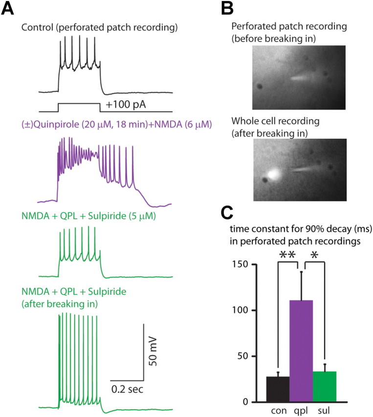
Quinpirole also induces an afterdepolarization during perforated-patch recordings from type A neurons. A, Recordings from a type A neuron in perforated-patch configuration (top three recordings) showing the quinpirole-induced afterdepolarization that occurs in the presence of NMDA, and is reversed by sulpiride. Bottom, shows a recording from the same neuron after breaking in and shifting to a whole-cell recording. B, Fluorescent dye in the recording pipette was excluded from the neuron while in the perforated-patch configuration (top), but entered the neuron after breaking in and shifting to a whole-cell configuration (bottom). C, Summary data showing that (±)quinpirole (20 μm) plus NMDA (6 μm) prolongs the time constant for the membrane potential to return to baseline following depolarizing current pulses (350 pA, 250 ms), and that this is reversed by the addition of sulpiride (5 μm) (n = 5). *p < 0.05, **p < 0.01.
