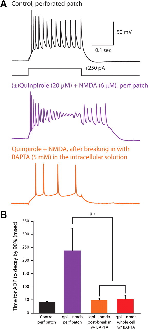
Figure 8.

The Ca2+ chelator BAPTA eliminates the quinpirole-induced afterdepolarization. A, Perforated patch recording from a type A pyramidal neuron in control conditions (top) and after application of quinpirole + NMDA elicits an afterdepolarization (middle). Bottom, In the same neuron, after breaking in and switching to a whole-cell recording configuration, the quinpirole-induced afterdepolarization is abolished. BAPTA (5 mm) is present in the pipette solution. B, Summary data showing time constants for the membrane potential to return to baseline following depolarizing current pulses (50–150 pA, 250 ms) under various conditions. “Control,” perforated patch recordings in control ACSF (black; n = 3); “qpl + NMDA, perf patch,” the same perforated patch recordings after applying quinpirole and NMDA (orange; n = 3); “qpl + NMDA, postbreak in w/BAPTA,” whole-cell recordings from the same cells that were initially recorded in perforated patch configuration (in quinpirole and NMDA) (purple; n = 3); “qpl + NMDA, whole-cell w/BAPTA,” recordings from cells that broke in and switched to whole-cell configuration during the application of quinpirole + NMDA (red; n = 5). **p < 0.01.
