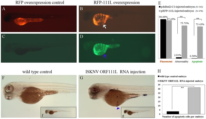
Figure 3. ISKNV ORF111L overexpression resulted in evident apoptosis in zebrafish embryo.
(A–B) The RFP and RFP-111L expression in pdsRed2-C1- and pRFP-111L-injected embryos were shown. (C–D) No obvious apoptotic signal was found in RFP overexpressing embryo, while increased apoptosis signal were clearly observed in RFP-111L-overexpressing embryo. (E) Statistics analysis of fluorescent, abnormality and apoptosis of embryos. Embryos shown above were at 3 dpf stage and represented the typical phenotype in three individual microinjection experiments. The significance of differences are calculated by the t-test (** indicates p<0.01). (F–G) TUNEL assay was performed in wild type and ISKNV ORF111L mRNA-injected embryos. Apoptotic cells were shown (arrow head). Figure F and G is the enlarged figure from panel f and g, respectively. (H) After the NBT/BCIP staining, the number of apoptotic cells was counted in wild type and ISKNV ORF111L mRNA-injected embryos.

