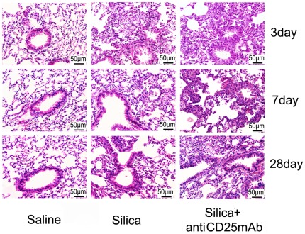
Figure 1. Histopathology changes in mouse lungs after instillation with HE staining (×200).
The scale on the graph above was 50 µm; date was day3, day7, day28. Lung sections were stained with H&E. The degree of inflammation was assessed by the histological analysis of six random fields per sample (with n = 5 mice per group).

