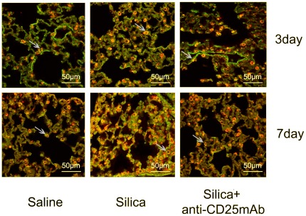
Figure 4. The localization of CD4+IL-17A+ cells in lung tissue examined by immunofluorescence (×600).
The CD4+(red) cells, IL-17A+(green) cells and CD4+IL-17A+(yellow) cells were colocalized in the lung tissue sections by confocal immunofluorescence microscope, analyzed with the leica confocal software package. Results from one representative experiment out of 5 (with n = 5 mice per group) are shown.

