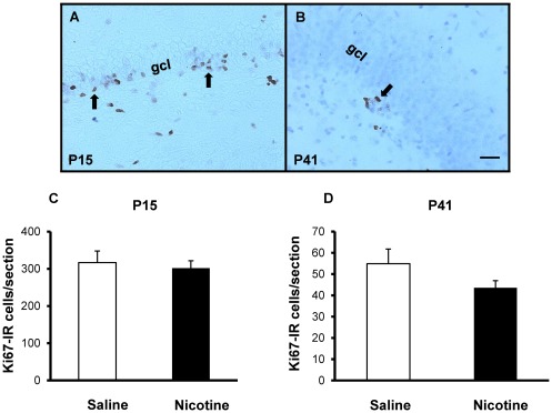
Figure 4. Cell proliferation.
DG proliferation assessed from Ki67-immunostained brain sections from juvenile (P15) and adolescent (P41) male offspring exposed to saline (controls) or nicotine throughout prenatal and postnatal development. Representative micrographs of Ki67-IR cells (arrows) in the subgranular zone or adjacent granule cell layer (gcl) in control P15 (A) and P41 (B) offspring. No significant difference in numbers of Ki67-IR cells was found in the dorsal hippocampus of P15 (C; p = 0.70) or P41 (D; p = 0.17) offspring. Scale bar = 25 µm.

