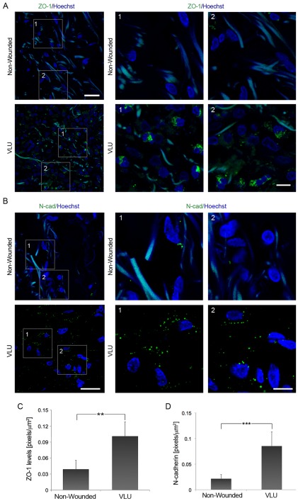
Figure 2. Increased expression of N-cadherin and ZO-1 in the dermis of human chronic VLU (A) ZO-1 expression levels are elevated in the dermis of chronic VLU compared to matched non-wounded controls (n = 6).
Scale bar = 25 µm. Higher magnifications of VLU and intact skin (boxed regions 1 and 2) stained for ZO-1 (green) and Hoechst (blue) are shown. Scale bar = 10 µm. (B) N-cadherin is significantly upregulated in chronic VLU compared to matched non-wounded samples (n = 6). Scale bar = 25 µm. The boxed regions 1 and 2 show high magnifications of VLU and non-wounded skin samples stained for N-cadherin (green) and Hoechst (blue). Scale bar = 10 µm. Values represent mean ± SD. (C and D) Graphs show ZO-1 and N-cadherin expression levels in VLU vs. non-wounded skin. Values for ZO-1 and N-cadherin were expressed as mean ± SD; **p<0.01 and p<0.005, respectively.

