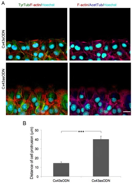
Figure 6. Targeting Cx43 induces cytoskeletal changes in leading-edge fibroblasts.
(A) Representative images showing the distribution of F-actin (red), and tyrosinated (green) and acetylated (blue) tubulin (TyrTub and AcetTub, respectively) in leading edge Cx43sODN and C43asODN fibroblasts, 3 h after wounding. Cells were also counterstained with the nuclear marker Hoechst. Scale bar = 20 µm. (B) The graph shows the length of the protrusions of wound edge cells. Data represent the distance (mean ± SEM) from the nucleus to the leading edge in n = 3 experiments (***P<0.005).

