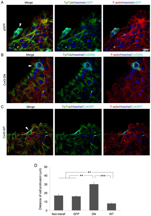
Figure 7. Modulation of Cx43 levels influence wound-edge cytoskeletal architecture in fibroblasts.
Representative images of leading-edge cells transfected (arrowhead) with (A) a control pGFP construct (B) Cx43-DN, or (C) Cx43-WT, stained for TyrTub, F-actin and Hoechst. Scale bar = 20 µm. (D) Graph depicting the length of lamelipodial protrusions of fibroblasts transfected with the different constructs is shown (**p<0.01; ***P<0.005; n = 3).

