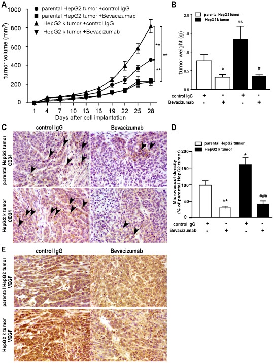
Figure 5. Bevacizumab impaired the tumor growth and angiogenesis in tumor-bearing mice in vivo.
2×10⁶ HepG2 k or parental HepG2 cells in cells in 200 µl phosphate buffered saline were injected by subcutaneous injection to obtain s. c. tumors. (A) Average tumor volume is shown for the HepG2 tumors. Parental HepG2 tumor with control IgG (n = 5), Parental HepG2 tumor with Bevacizumab (n = 5), HepG2 k tumor with control IgG (n = 5) and HepG2 k tumor with Bevacizumab (n = 5). **, P<0.01. (B) Mice were killed after 28 days implantation and the tumor tissues were removed and weighed. *, P<0.05. (C–D) Twenty-eight days after implantation, the numbers of new microvessels marked with CD34 (arrow heads) in the subcutaneous tumors were quantified by performing new vessel counts of 10 random fields at ×400 magnification. *, P<0.05, **, P<0.01 versus parental HepG2 tumor respectively; ###, P<0.001 versus HepG2 k tumor. (E) Immunohistochemistry analysis of the expression of VEGF in implanted tumors.

