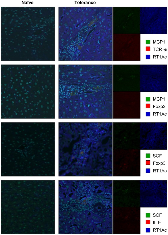
Figure 2. Colocalization of recipient-derived hepatic mast cells, Tregs (Foxp3+/TCR γδ chain+) and cytokines (SCF and IL-9) for mast cell activation.
Cryosections of naïve and OLT livers at tolerogenic phase (>60 days) were immunoprobed with goat polyclonal Ab against MCP1, mouse monoclonal Ab against γδ TCR, Foxp3 or IL-9, rabbit polyclonal Ab against SCF, and rat polyclonal Ab against RT1Ac (specific to PVG recipients) followed by incubation with Alexa Fluor® 488−conjugated donkey anti-goat or rabbit IgG, HyLite Fluor™ 594−conjugated goat anti-mouse IgG or Alexa Fluor® 647−conjugated goat anti-rat IgG. Hoechst 33342 (specific to nucleus) was used for counterstaining. Data are representative of six individual liver sections (60× magnification). Right columns indicate the data without merging.

