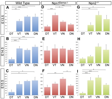
Figure 3. Loss of Npn1 signaling through Sema disrupts the pattern of innervation of the corneal quadrants.
Whole mount corneas were immunostained with TuJ1 and imaged. The lengths of nerve bundles projecting into each quadrant were quantified as described in the Methods section. For all graphs, Y-axis is average length of corneal nerves (µm) and X-axis is cornea quadrants. (A–C) Wild type, (D–F) Npn1Sema−/− mutant, and (G–I) Npn2−/− mutant eyes were analyzed at E13.5–15.5. ANOVA with a Tukey post test was performed on all data sets. For all samples n = 8, except Npn2−/− mutants where n = 6 for E14.5 and E15.5. No bracket indicates P>0.05; *, P<0.05; **, P<0.01; ***, P<0.001.

