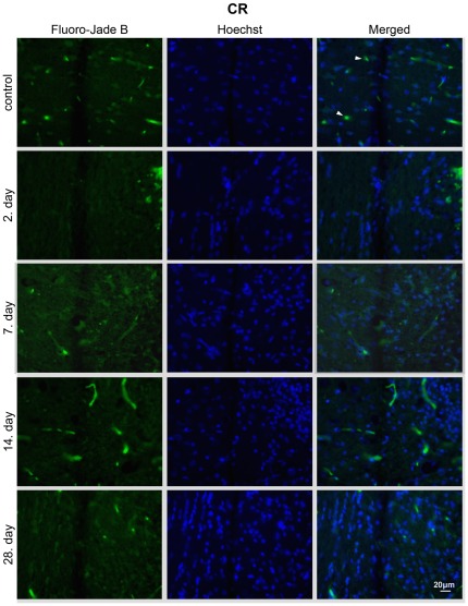
Figure 5. CR treatment suppresses neuroapoptosis in the perilesioned area.
Fluoro-Jade B and Hoechst staining of brain sections from CR animals 2, 7, 14, 28 – days after injury. Fluoro-Jade B/Hoechst positive cells were not detected in the CR group at any time point. Images are representative of brain sections at the site of the lesion (n = 3 animals per experimental group); c – physiological control; 2, 7, 14, 28 – days after the injury; blood vessels (arrowheads); magnification 40×.

