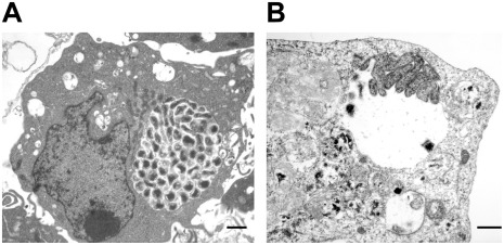
Figure 2. Variations of morulae in infected macrophages and tick cells.
E. chaffeensis containing phagosomes within the infected macrophages (A) were more compact with organisms occupying most of the intra-morulae space. The organisms in infected tick cells (B) were mostly aggregated at one end of the morula or attached to the morula membrane, intra-morulae space is also considerable more in the tick cell phagosomes and the morula size is also larger. (Scale bar 1 µm).

