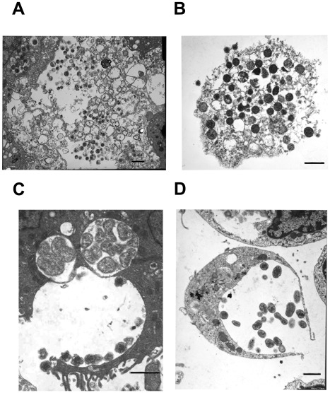
Figure 8. Release of E. chaffeensis from infected macrophages and tick cells.
Most of the infected host cells exhibited release by complete lysis. A subset of the infected cells also released organisms by exocytosis by creating an opening to the morula membrane. (A and C, infected macrophages and B and D, infected tick cells) (Scale bar 1 µm).

