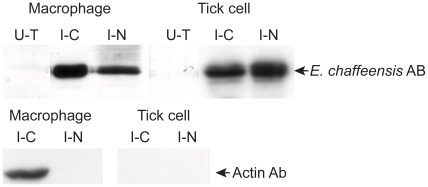
Figure 12. Western blot analysis to identify E. chaffeensis proteins.
Total cell lysates from uninfected cells, cytoplasm (C) and nucleic (N) fractions from E. chaffeensis-infected macrophages and tick cells were assessed by immunoblot analysis using E. chaffeensis mAb 56.5 that recognizes p28 Omp 19 [22]. E. chaffeensis infected macrophage and tick cell protein fractions were also probed with β actin Ab. (U–T, uninfected cell-derived total soluble proteins; I–C, E. chaffeensis-infected cell derived cytoplasmic proteins; I–N, E. chaffeensis-infected cell derived nucleic proteins).

