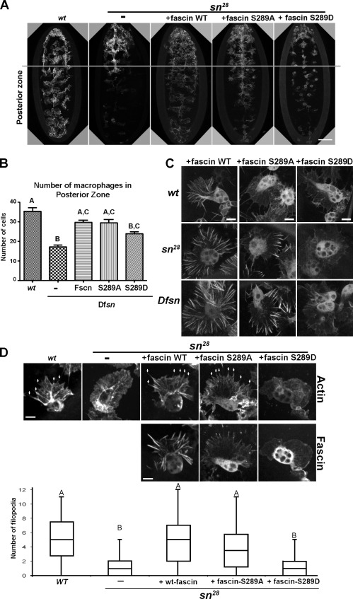
Figure 1.
Drosophila fascin S289 regulates embryonic macrophage migration. (A) Comparison of macrophage dispersal on the ventral surface of stage 15 embryos in wild-type (wt) and deficiency (Dfsn) mutants expressing GFP-fascin transgenes. The gray line demarcates the anterior one third of the embryo. (B) Bar graph revealing the mean number of macrophages that migrated from the anterior region of the embryo (n = 10 embryos). Letters highlight no significant difference by one-way ANOVA (P < 0.05). Error bars show means ± SD. (C) Wild-type GFP-fascin and GFP-fascin S289A/D were expressed specifically in macrophages in wild-type and singed mutant embryos (sn28 and Dfsn). In the fascin mutants, fascin S289A localized to short filaments at the distal region of lamellae, whereas S289D remained cytoplasmic. (D) Filopodia (arrows) were quantified in wild type, the singed mutant, and singed mutants expressing fascin transgenes using a fluorescent actin probe. Letters highlight no significant difference by one-way ANOVA (P < 0.05). Boxes define the 25th and 75th percentile. The bands in the middle are the medians. The whiskers represent the 1.5 interquartile range. Bars: (A) 50 µm; (C and D) 5 µm. See also Video 1.

