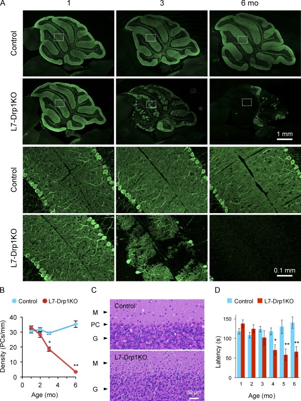
Figure 1.
Purkinje cells degenerate in L7-Drp1KO mice. (A) Immunofluorescence of cerebellar sagittal sections around the median line using antibodies to Car8, a Purkinje neuron marker. Control and L7-Drp1KO mice were analyzed at the indicated ages. Boxed regions show magnified images. (B) Quantification of Purkinje cell density. The number of soma of Purkinje cells was determined and normalized relative to the length of the Purkinje cell layer. Values represent the mean ± SEM (n ≥ 3). (C) Hematoxylin and eosin stains of cerebellar sagittal sections at 6 mo. The Purkinje cell layer (PC) was lost in L7-Drp1KO mice. M, molecular layer; G, granule cell layer. (D) Rotarod tests. To examine motor coordination ability, the latency to fall from the rotarod was determined in control and L7-Drp1KO mice. Values represent the mean ± SEM (n ≥ 5).

