Figure 10.
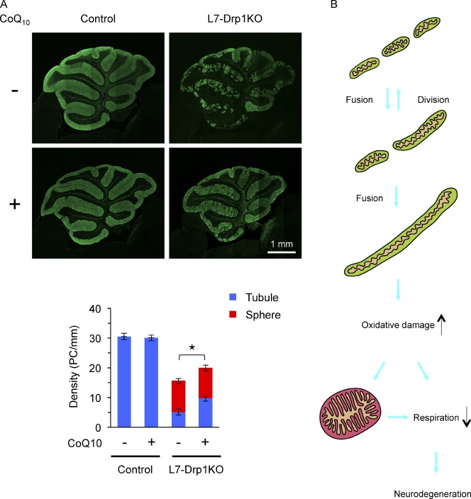
Coenzyme Q10 suppresses Purkinje cell degeneration in L7-Drp1KO mice. (A) Control and L7-Drp1KO mice were fed control and 1% coenzyme Q10–containing diets from 3 wk to 3 mo of age and then examined by immunofluorescence using antibodies specific to Car8 and PDH. Shown are cerebellar sagittal sections around the median line that were stained by the anti-Car8 antibodies. The number of soma of Purkinje cells (PC) that contained tubular (blue) and swollen (red) mitochondria was determined and normalized relative to the length of the Purkinje cell layer. Values represent the mean ± SEM (n ≥ 5). (B) A model for neurodegeneration caused by mitochondrial division deficiency. In wild-type cells, mitochondria fuse and divide, as well as maintain a short tubular morphology. When mitochondrial division is blocked, the organelles elongate due to imbalanced, excess fusion. Elongated tubules accumulate oxidative damage, which leads to swelling of mitochondria and impairment of the electron transport chain. Mitochondrial enlargement may also decrease respiratory competence. Eventually, this decline in respiration causes neuronal death.

