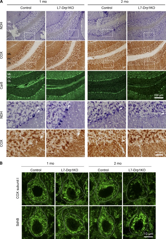
Figure 3.
Decreased respiratory activities in L7-Drp1KO Purkinje cells. (A) Fresh frozen cerebellar sections of control and L7-Drp1KO mice at the indicated ages were stained for NADH dehydrogenase activity, COX activity, and Car8. Sagittal sections of the cerebellum around the median line were used. Boxed regions show magnified images. PC, Purkinje cell layer; M, molecular layer; G, granule cell layer. (B) Sagittal sections of the cerebellum in control and L7-Drp1KO mice at the indicated ages were stained using antibodies against Car8, cytochrome c oxidase subunit I (COX subunit I), or succinate dehydrogenase iron-sulfur subunit (SdhB). The soma of Purkinje cells are outlined based on Ca8 staining (dotted line).

