Figure 6.
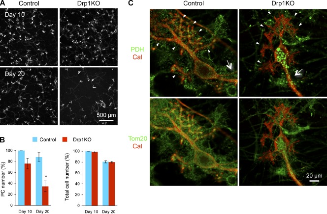
Purkinje cells die after Drp1 loss in vitro. (A) Cerebellar neurons were isolated from P0 Drp1flox/flox mice and cultured for 20 d. When cells were plated (d 0), lentiviruses carrying Cre recombinase and GFP in tandem (Drp1KO) or GFP alone (control) were added to the culture. At d 10 and 20, cells were fixed and analyzed by immunofluorescence using anti-calbindin antibodies. (B) Quantification of the number of Purkinje cells and total cells. To count the number of Purkinje cells and total cells, calbindin staining and DAPI staining were used, respectively. Cell numbers were normalized to those of control cells at d 10 in each experiment. Values represent the mean ± SEM (n = 3). (C) Cultured cerebellar neurons were infected with lentiviruses carrying the Cre recombinase (Drp1KO) and the myc epitope (control). Mitochondria were visualized with immunofluorescence using antibodies to PDH, calbindin, and Tom20 in control and Drp1KO Purkinje cells at d 10. Dendrites and axons are indicated by arrowheads and arrows, respectively.

