Figure 9.
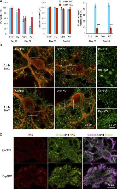
N-acetylcysteine suppresses mitochondrial enlargement, cell death, and oxidative damage in Drp1KO Purkinje cells. (A) Cerebellar neurons were isolated from P0 Drp1flox/flox mice and cultured for 30 d. At d 7, lentiviruses carrying the Cre recombinase (Drp1KO) or the myc epitope (control) were added to the culture medium. At the same time, cells were left untreated or treated with 1 mM N-acetylcysteine. At d 20 and 30, cells were fixed and analyzed by immunofluorescence using anti-calbindin antibodies. The number of Purkinje cells was quantified (left graph). DAPI staining was used to determine the total cell number (middle graph), which was normalized to that of untreated control cells at d 20 in each experiment. Purkinje cells that contained enlarged mitochondria were quantified (right graph). Values represent the mean ± SEM (n = 4). (B) Immunofluorescence of control and Drp1KO Purkinje cells was performed with antibodies against PDH (green) and calbindin (red) at d 20. Boxed regions show magnified images of mitochondria. (C) Immunofluorescence of control and Drp1KO Purkinje cells using antibodies to 4-hydroxynonenal (HNE), Tom20, and calbindin at d 30.

