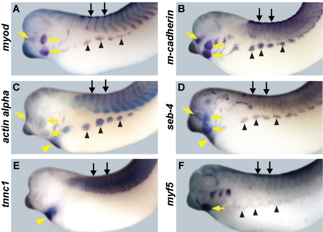
Figure 4. Expression patterns of muscle target genes.
In stage 35–37 embryos myod, m-cadherin, actin alpha, seb-4, tnnc1 and myf5 are all expressed in skeletal muscle including somites (black arrows), migrating hypaxial muscle anlagen (black arrowheads) and jaw muscle (yellow arrows). myod, m-cadherin, actin alpha, seb-4, and myf5 (A-D and F) are expressed in the somites, migrating hypaxial muscle anlagen and jaw muscle, and these expression patterns overlap with those of ebf2 and ebf3 (Figure 2). m-cadherin (B) is weakly expressed in a central band in somites, with expression throughout the somite. myf5 (F) expression in somites is weaker than other genes at this stage, and is expressed at the leading edge of migrating hypaxial muscle. tnnc1 (E) is expressed in the somites. actin alpha, seb-4, and tnnc1 are expressed in the heart (yellow arrowheads). All embryos show lateral views.

