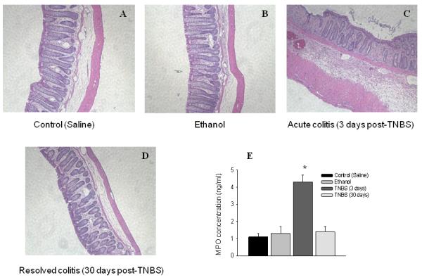
Figure 1. Histological and biochemical evaluation of the severity of colonic inflammation induced by TNBS.

Hematoxylin and eosin (H&E) staining of the distal colon from the following groups of animals: control (A, saline), ethanol (B, 3 days), acute colitis (C, TNBS - 3 days) and resolved colitis (D, TNBS - 30 days). Please note that only group with acute colitis (3 days post-TNBS) shows significant tissue damage including sites of infiltration, thickening of the muscle layer, and disruption of the colonic crypts. E, Concentration of myeloperoxidase (MPO) enzyme in the distal colon from control and experimental groups correlates with the results of histological evaluation. *P≤ 0.05 to control group.
