
Figure 3.
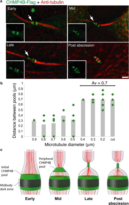
ESCRT-III fission complex originates at the location of the initial ESCRT-III ring and migrates to a conserved distance. Synchronized MDCK cells expressing CHMP4B-Flag were stained with anti-α-tubulin (red) and anti-Flag (green) antibodies. (a) Images show typical localization patterns observed for CHMP4B at intercellular bridges of cells undergoing abscission. The time course of abscission (early – postabscission) was determined based on the microtubule diameter measured on the side of the intercellular bridge that is undergoing constriction (see arrow). (b) Distribution of the distances measured between the initial and peripheral CHMP4B pools as a function of microtubule diameter. The distance between the CHMP4B pools was measured on either side of the intercellular bridge, when applicable, and correlated to the minimum value of microtubule diameter measured on that side. When a continuous elongated initial pool of CHMP4B was observed (as in a early) the distance between the pools was given a 0 value. Distances smaller than 0.2 μm were not measured due to the resolution limit of confocal microscopy and were given a 0 value. Average values for each microtubule diameter are shown as columns. Notably, the distance between the two ESCRT-III pools stabilizes in intercellular bridges with a diameter of 0.4 μm or smaller. The average distance determined for these diameters was 0.7 ± 0.1 μm. n = 26. (c) Schematic representation for ESCRT-III distribution (green) at the intercellular bridge during cytokinetic abscission. Microtubules are labeled in red.