
Figure 4.
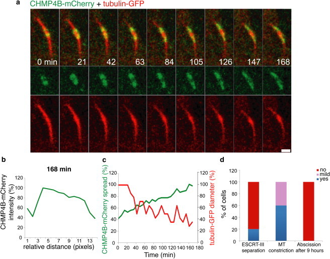
Separation of the central ESCRT-III complex into two distinct complexes is ATP dependent. Live MDCK cells expressing CHMP4B-mCherry (green) and tubulin-GFP (red) were subjected to acute ATP depletion (as described in Materials and Methods) and imaged during cytokinesis at 7 min intervals. (a) Representative images of every third frame from the movie sequence are shown. The overlay image is shown in the upper panel and the individual channels are shown below. (b) Line intensity profile of CHMP4B-mCherry on one side of the intercellular bridge at t = 168 min showing the continuity of the signal along the intercellular bridge and the lack of the characteristic two distinct ESCRT-III pools described in Fig. 2, b–d. (c) Measurements of the relative spread of CHMP4B-mCherry signal along one side of the intercellular bridge (green) and the relative microtubule diameter measured on this side (red) (see Materials and Methods). Shown is a representative example of 10 independent experiments. A summary of all the experiments conducted and the parameters evaluated is shown in d. Scale bar = 2 μm.