
Figure 5.
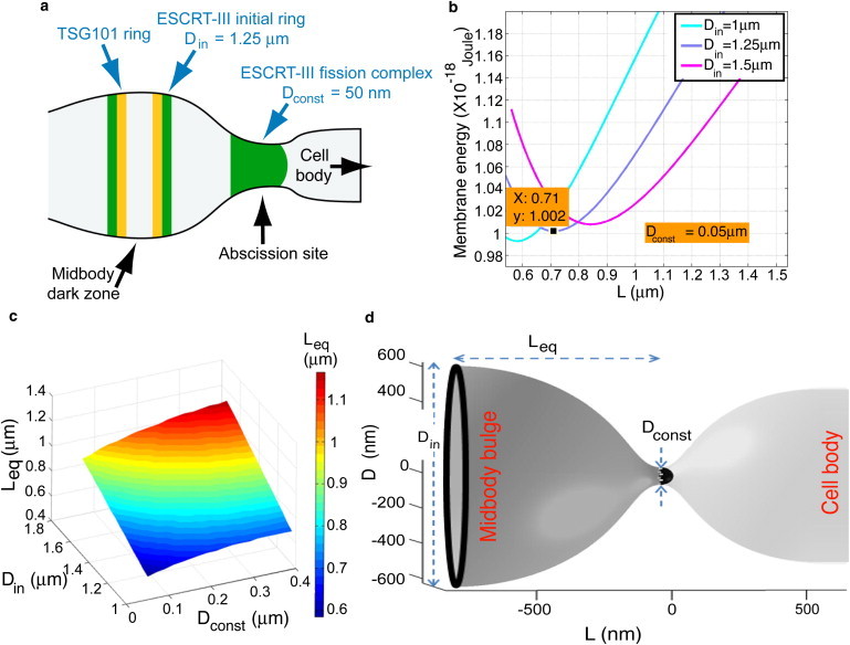
Computational analysis of the equilibrium distance between the two ESCRT-III complexes. (a) Schematic representation of the localization of ESCRT components in intercellular bridges of cells considered in our model. The initial diameter of ESCRT-III (Din) is based on our previous direct measurements of the diameter of CHMP4B rings located at the edge of the midbody dark zone (6). The diameter of the ESCRT-III peripheral pool in the abscission site (Dconst) is taken as the spontaneous diameter measured for the ESCRT-III complex in vitro (8). ESCRT-I is depicted in yellow. ESCRT-III is depicted in green. (b) The bending elastic energy of the membrane between the two ESCRT-III pools as a function of the distance between the ESCRT-III pools. The three curves correspond to different diameters of the initial pool. Din = 1 (light blue), 1.25 (purple), 1.5 μm (magenta), whereas the diameter of the narrow constriction pool is taken Dconst = 0.05 μm. The curves show the energy minimum corresponding to the equilibrium distance Leq. (c) The equilibrium distance, Leq, as a function of the diameter of the initial pool, Din, and the diameter of the peripheral pool, Dconst. (d) A representative computed shape of the lipid membrane between the two ESCRT-III pools: the initial pool at the edge of the midbody dark zone and the peripheral pool at the constriction site. The computed distance between the pools corresponded to Din = 1.25 μm and was Leq = 0.71 μm (see graph in b). The constriction diameter in this frame is taken to be Dconst = 0.1 μm for better visualization.