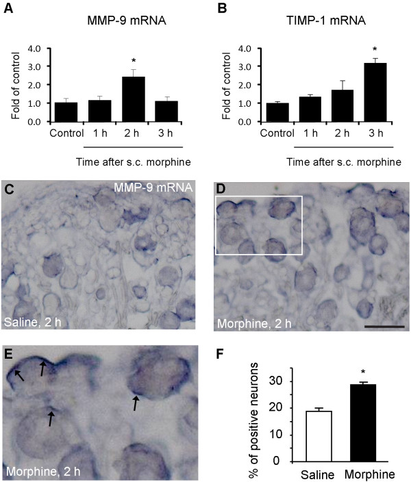
Figure 2.
Subcutaneous morphine induces MMP-9 and TIMP-1 mRNA up-regulation in DRGs. (A, B) RT-PCR showing time course of subcutaneous morphine-induced MMP-9 (A) and TIMP-1 (B) mRNA expression in DRGs. *P < 0.05, compared to naive control, ANOVA followed by Bonferroni post hoc test, n = 4 mice. (C-E) In situ hybridization showing MMP-9 mRNA expression in DRG neurons 2 h after subcutaneous injection of saline (A) and morphine (B, C). Scale, 50 μm. The box in D is enlarged in E. Note strong MMP-9 mRNA staining (arrows) near cell surface following morphine treatment. (F) Percentage of MMP-9 mRNA-positive neurons in DRGs. *P < 0.05, compared to vehicle (saline), Student's t-test, n = 4 mice.

