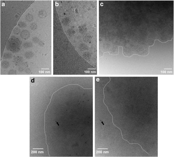
Figure 4.
Cryo-electron microscopy shows intact virions after INA treatment and recognizable hemagglutinin epitopes following Triton treatment. Influenza (PR8) suspensions were treated with INA + UVA for 30 minutes, then treated with 0.5% Triton X-100 for 1 hour, then fixed and frozen for cryo-EM. (a) control influenza, (b)influenza + INA + UVA for 30 minutes, (c) "b" plus Triton, (d)immunogold labeling of virus + INA + UVA + Triton, (e) Control immunogold sample same as "d" without primary antibody to assess non-specific binding of secondary immunogold. White lines are drawn to guide the eye in panels "c", "d" and "e", indicating the approximate boundary between the aggregated viral proteins and the bare grid. Arrows indicate immunogold location, in panel "d" within the aggregate, and panel "e" outside the aggregate. Images shown are representative of sections of overall grid.

