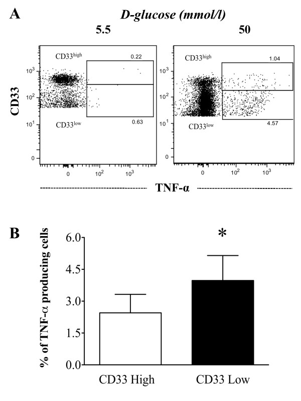
Figure 5.
TNF-α production in CD33lowand CD33highmonocytes. Monocytes from healthy donors were cultured in the presence of glucose for 7 days and were then stained with anti-human CD33 and anti-human TNF-α antibodies. (A) Representative dot-plots show the percentages of TNF-α-producing CD33low and CD33high monocytes that were cultured with either 5.5 mmol/l (left) or 50 mmol/l (right) D-glucose (B) The bar graph summarizes the levels of TNF-α production by CD33low (white) and CD33high (black) monocytes cultured with 50 mmol/l D-glucose (n = 4). The data are expressed as the mean ± SD. * P < 0.05 as compared to the TNF-α production by CD33low and CD33high

