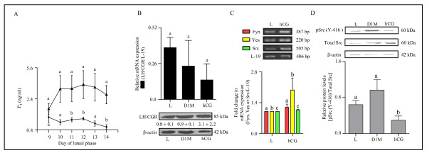
Figure 6.
Expression and characterization of LH/CGR and genes belonging to SFKs in the monkey CL during simulated early pregnancy condition. (A) The graph illustrates circulating mean P4 levels during different days of the luteal phase without (solid circles) and with hCG (solid triangle) treatment. Each point represents mean ± SEM values (n = 3 animals/control or hCG treatment). Bars with different letters indicate statistical significance (p < 0.05). (B) The qPCR expression for LH/CGR mRNA (upper panel) and the levels of LH/CGR protein (lower panel) in the CL from monkeys collected on day 14 without treatment i.e., late CL (L), on day 1 of menses (D1M) and on day 14 following hCG treatment from day 9-13 of luteal phase (hCG). Each bar in the upper panel represents mean ± SEM values (n = 3 animals/treatment). For comparison among various groups, one-way ANOVA analysis was performed and the data across groups was not significantly different (p > 0.05). The representative immunoblots probed with anti-LH/CGR antibody and anti-β-actin antibody are shown in the lower panel. Densitometric analysis (mean ± SEM for n = 3) of immunoblots was performed and the protein levels are indicated below respective bands. L19 mRNA was used as internal control for qPCR, while β-actin protein level was used as loading control. (C) Semi-quantitative RT-PCR expression of genes belonging to SFKs (Fyn, Yes and Src) in the CL collected from monkeys on day 14 of luteal phase that received hCG treatment to stimulate early pregnancy (hCG) or without treatment Late (L). Each bar represents mean ± SEM values (n = 3 animals/control or hCG treatment). L-19 mRNA was used as internal control and fold change in mRNA expression was calculated following densitometric analysis. (D) Immunoblot analysis was performed to determine functional activation of Src protein i.e., protein levels of active pSrc (Y-416) and total Src in the CL collected from monkeys on late luteal phase (L), on day 1 of menses (D1M) and on day 14 of luteal phase following hCG treatment on day 9-13 of luteal phase (hCG). A representative immunoblot for each of the protein antibody probed is shown along with the size of the protein. Anti-β-actin antibody (the protein loading control) probed blot is presented to indicate equal loading of protein in each lane. Densitometric analysis of immunoblots was determined and level of pSrc (Y-416) is expressed as mean ± SEM relative to the intensity of total Src/β-actin. Individual bars with different letters are significantly different (p < 0.05).

