Figure 1.
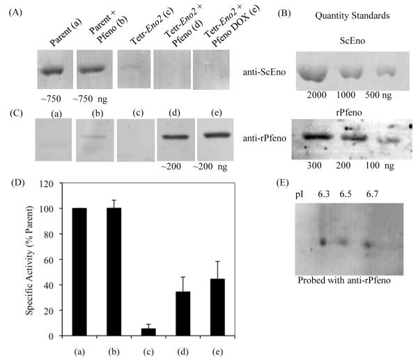
Quantification of enolase in (a) Parent strain, (b) Parent strain transformed with P. falciparum enolase (Parent + Pfeno), (c) Tetr-Eno2 strain, (d) Tetr-Eno2 strain transformed with Pfeno and grown in the absence and (e) in the presence of 10 μg/mL doxycycline (DOX). Western blot analysis was performed for the quantification of the expressed enolase (ScEno or rPfeno) in cell extracts of different strains of yeast. (A) Western blot of yeast enolase; (B) Western blot of pure ScEno or rPfeno used as standards; (C) western blot of Pfeno. Equivalent amounts of protein were loaded in each lane. (D) Specific activity of enolase in various cell extracts plotted as the percent of the parent strain and (E) Western blot of two dimensional gel electrophoresis showing the variant profile of Pfeno in Tetr-Eno2 + Pfeno (DOX) yeast cells. Anti-rPfeno antibody was used to probe the membrane.

