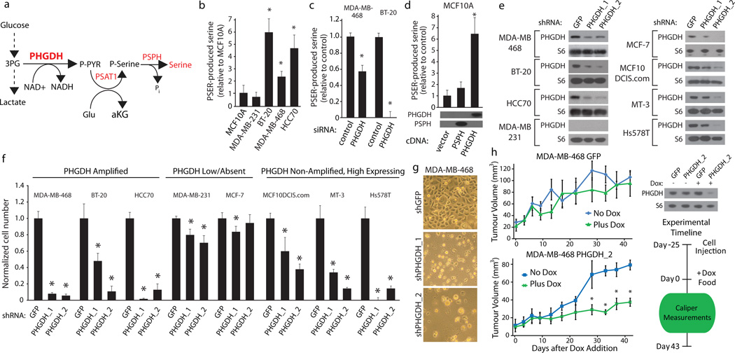
Figure 3. Cell lines with elevated PHGDH expression have increased serine biosynthetic pathway activity and are sensitive to PHGDH suppression.
a, Serine biosynthesis pathway (SBP). b–d, Serine production by SBP in (b) indicated breast cell lines, (c) after PHGDH suppression by siRNA, and (d) MCF-10A cells expressing PHGDH or PSPH cDNAs with associated immunoblots. e–f, Immunoblots of indicated proteins (e) for indicated cell lines expressing control shRNA (GFP) or shRNAs against PHGDH (PHGDH_1 and PHGDH_2). Relative proliferation (f) of cells transduced with shRNA constructs after seven days. g, Images showing cellular morphology of MDA-MB-468 at day seven of (f). h, Tumour growth of MDA-MB-468 cells expressing doxycycline inducible control shRNA (GFP) or shRNA against PHGDH (shPHGDH_2) in mice fed doxycycline (Dox, 2mg/kg, green lines, n=5) or normal (blue lines, n=4) diet after initial tumour palpation (Day 0). Immunoblots of PHGDH or RPS6 (S6) shown for cells in vitro. Asterisks indicate p < 0.05 relative to control. Error bars for metabolite measurements (n=4) and tumour size indicate SEM and for cell number indicate SD (n=3).

