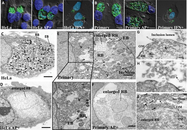
Fig. 2.
C. trachomatis infection in genital tract epithelial cells. (A, B) Morphological analysis by epifluorescent microscopy and deconvolution of C. trachomatis-infected cells unexposed or exposed to ampicillin (AP+) or IFN-γ (IFN+). Infected HeLa cells (A) and primary human endocervical epithelial cells (B) were fixed at 36 h p.i. MOMP was visualized as green. Host nuclei and bacteria were visualized as blue by DAPI staining. All images shown are overlays of fluorescence images (green and blue) on differential interference contrast (DIC) images (grayscale). Scale bars, 5 µm. (C, D) TEM of C. trachomatis inclusions reveals the presence of productive forms in the absence of ampicillin (C) and the accumulation of enlarged persistent forms in the presence of ampicillin (D) in C. trachomatis-infected HeLa cells. Scale bars, 2 µm. (E, F) TEM of C. trachomatis inclusions in infected primary human endocervical epithelial cells unexposed (E) or exposed to ampicillin (F). An enlarged image of multivesicular structures from (E) is shown below. V, multivesicular structures; IncMem, inclusion membrane (outlined with dotted line). Scale bars, 2 µm. (G–I) C. trachomatis produces OMVs in cultured primary human endocervical epithelial cells exposed to ampicillin as determined by TEM. OM/IM, outer/inner membrane; IncMem, inclusion membrane. Representative OMVs are indicated by arrows. Scale bars, 200 nm (G, H) or 100 nm (I).

