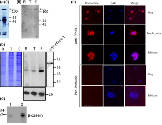
Fig. 1.
Expression of Pfnek-1 in asexual parasites. (a) Western blot analysis detecting the full-length protein. Asynchronous (i, chicken antibody) or synchronous (ii, rabbit antibody) parasite extract (15 µg) was loaded on the gels. Arrows indicate the position of Pfnek-1. (b) Western blot analysis (rabbit antibody) showing that the antibody recognizes recombinant GST-Pfnek-1 (Dorin et al., 2001) (far right lane) and displays a loading control (antibody against P. falciparum 2-Cys peroxiredoxin). R, Rings; T, trophozoites; S, schizonts. Molecular masses of co-migrating markers are indicated in kDa. (c) Immunofluorescence analysis. Synchronized asexual parasites were stained with the immunopurified rabbit anti-Pfnek-1 antibody (top panels). The two bottom panels display negative controls using pre-immune serum. Bar, 5 µm. (d) Immunoprecipitated kinase activity. The anti-Pfnek-1 rabbit antibody was used to immunoprecipitate β-casein kinase activity from 3D7 parasite extracts (lane 2). The pre-immune serum was used as a control (lane 1).

