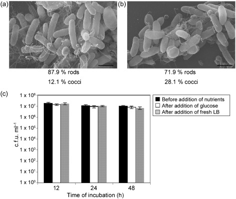
Fig. 5.
V. cholerae cells in coccoid morphology revert back to rod-shaped morphology with nutrient supplementation. (a) Electron micrograph of V. cholerae cells incubated for 12 h in CM50, given glucose to a final concentration of 1 %, and incubated for an additional hour at 30 °C. (b) Electron micrograph of V. cholerae cells incubated for 12 h in 2 ml CM50, given 2 ml fresh LB, and incubated for an additional hour at 30 °C. The number of bacteria in coccoid and rod morphology was quantified, and the percentages of these cells are given below each figure. Bars, 1 µm. (c) Quantification of cells grown on solid growth medium before and after the addition of nutrients. Prior to the addition of nutrients, the majority of cells were in coccoid morphology after 12 h, as shown in Figs 2 and 3. Results shown are mean and sd.

