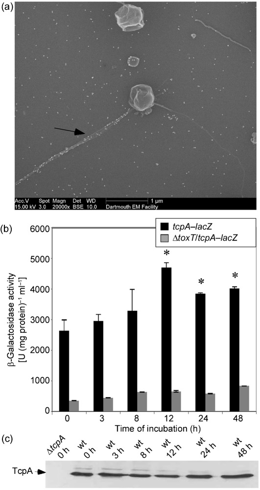
Fig. 6.
Expression of tcpA by V. cholerae in the coccoid form. (a) Electron micrograph of coccoid V. cholerae incubated for 12 h in CM50 and then immunolabelled with α-TcpA antibodies. The arrow indicates TCP. (b) β-Galactosidase activity of tcpA–lacZ and ΔtoxT/tcpA–lacZ (negative control). Expression of tcpA was found to significantly increase compared with the 0 h time point, as indicated by the asterisks (P<0.0002). Results shown are mean and sd. (c) Western blotting using α-TcpA6 peptide antibodies was performed on whole-cell samples at various time points after incubation in CM50.

