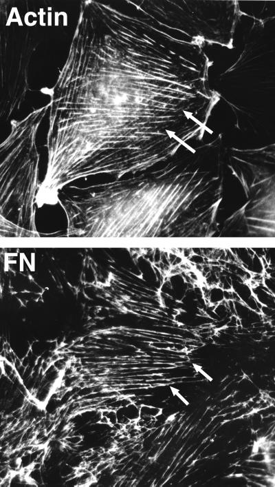
Figure 2.
Coalignment of actin microfilament bundles and extracellular FN fibrils. Double-label immunofluorescence of a well spread, nonmotile cell showing extensive codistribution of the two sets of fibrils. Note the extension beyond the cell boundary of FN fibrils linked to stress fibers within the cells (arrows). This demonstrates the tension applied by cells on the ECM. Reproduced with permission from Hynes and Destree, 1978 (4).

