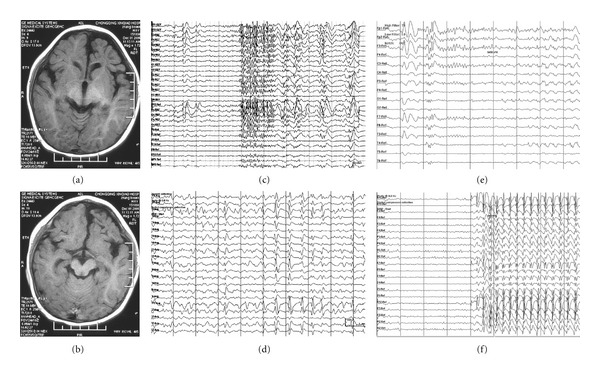
Figure 2.

Clinical data of a patient with left frontal lobe atrophy. (a) and (b): MRI scan showing atrophy of left frontal lobe. (c) EEG showing PFA. (d) EEG showing SSW. (e) EEG showing epileptiform discharges during a tonic seizure. (f) EEG showing epileptiform discharges during an atypical absence. Data were from patient no. 13 in Tables 1 and 2.
