Figure 7.

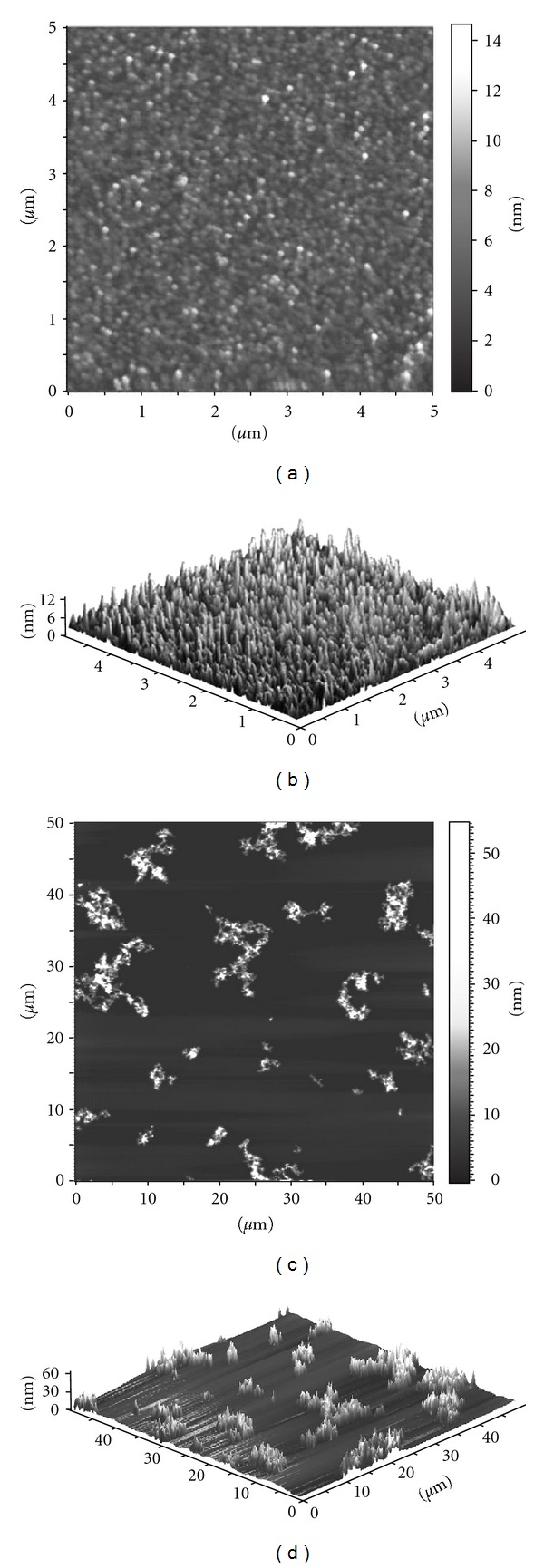
AFM images of anatase sample 3.3: 2D (a) and 3D (b) view of the freshly prepared acid sol; 2D (c) and 3D (d) view of the neutralized sol.

AFM images of anatase sample 3.3: 2D (a) and 3D (b) view of the freshly prepared acid sol; 2D (c) and 3D (d) view of the neutralized sol.