Figure 2.

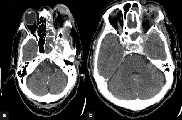
(a) CT scan of the orbit showing proptosis and distortion of the left globe (b) CT scan of brain (post contrast) showing an enhancement of the cavernous sinus suggestive of thrombosis

(a) CT scan of the orbit showing proptosis and distortion of the left globe (b) CT scan of brain (post contrast) showing an enhancement of the cavernous sinus suggestive of thrombosis